


Cardiological examinations
Regular preventive check-ups help to detect cardiovascular diseases early.
Cardiovascular diseases are among the most common causes of death in Germany. However, many risk factors responsible for cardiovascular disease can be avoided or detected early through preventive measures.
In Germany, preventive examinations (known as "check-ups") are covered by statutory health insurance every three years from the age of 35.
Typical risk factors for heart diseases include high blood pressure, elevated LDL cholesterol, smoking, diabetes, lack of physical activity, obesity, and a family predisposition.
The following symptoms can be initial signs of a cardiovascular disease:
- Shortness of breath during light physical activity
- Dizziness
- Fainting spells
- Pressure or pain in the chest
What cardiological examination methods do we offer?
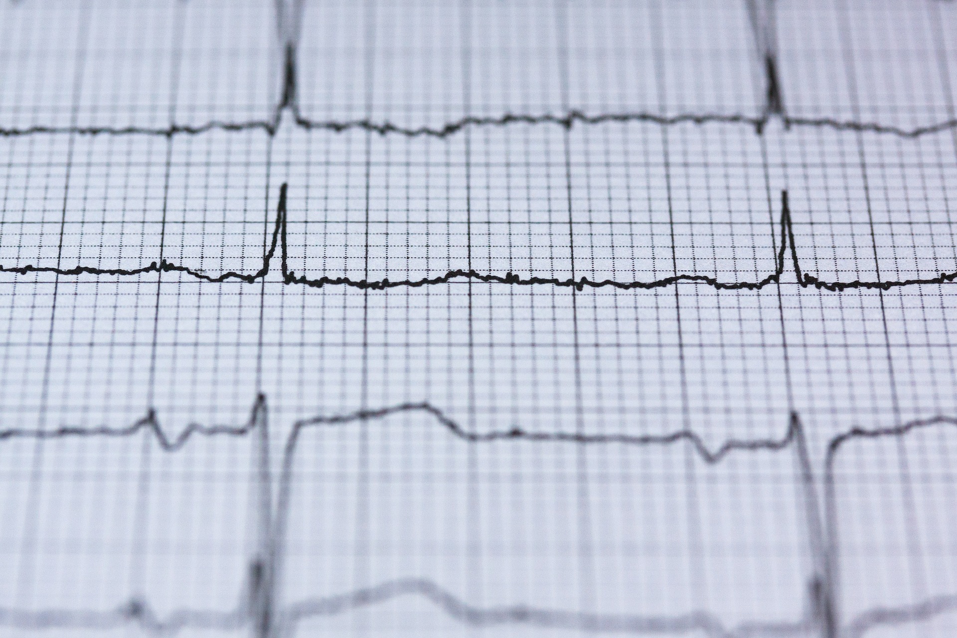
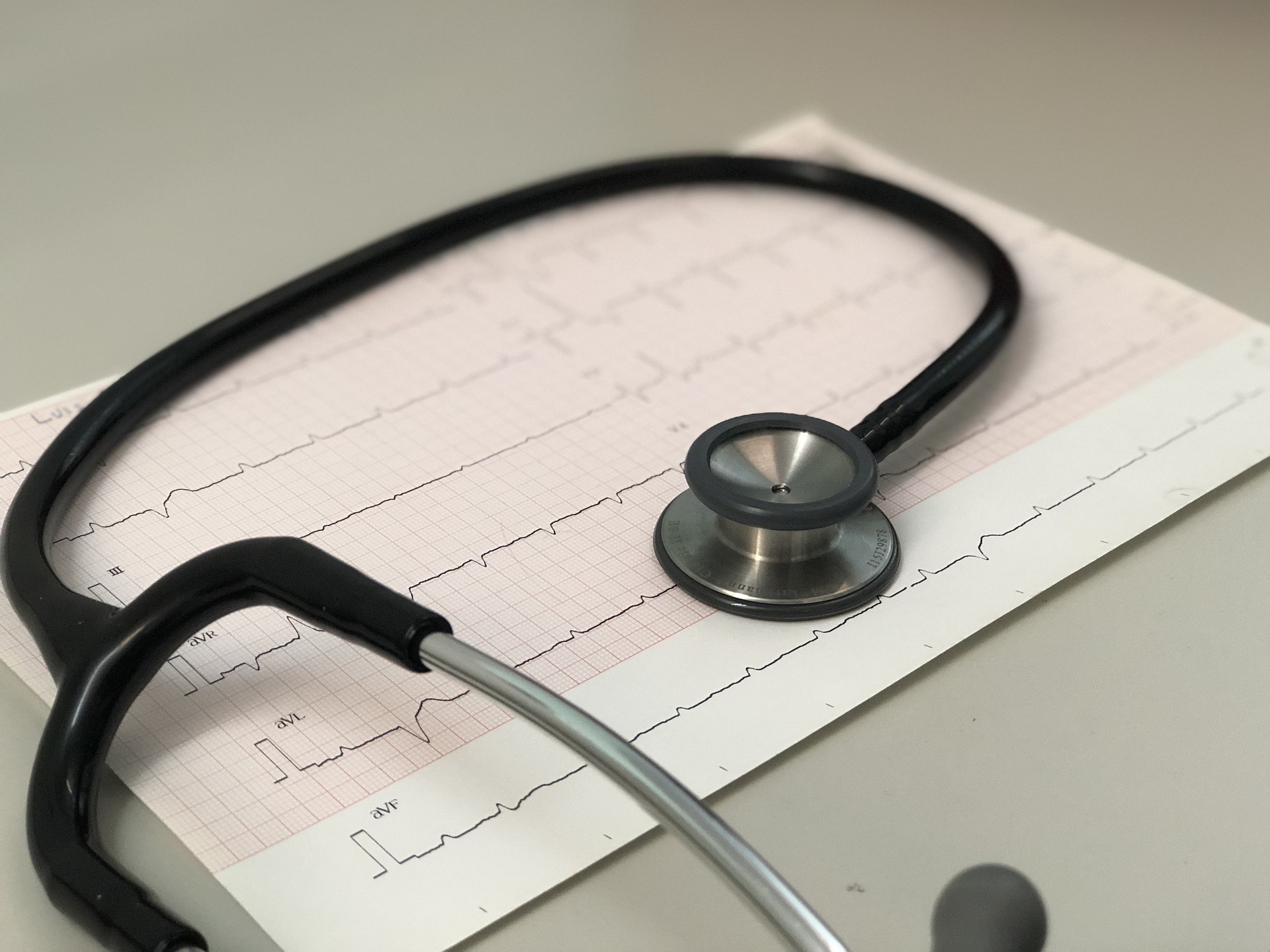
ECG
An electrocardiogram (ECG) records electrical currents generated by heart actions. These voltage changes are measured using a total of 12 electrodes that are attached to the chest wall, arms, and legs of the lying patient. The so-called 12-lead ECG can provide indications of a heart attack or rhythm disorders, among other things.An ECG examination usually lasts only a few minutes and is completely painless and harmless.
Stress-ECG ➤
The stress-ECG records heart activity under constant monitoring of blood pressure during increasing physical activity. For this purpose, we use a bicycle ergometer. The patient has electrodes attached to their body that record the electrical currents generated by heart actions.
The stress-ECG is used, for example, to detect circulatory disorders in certain areas of the heart and to record blood pressure behavior or the occurrence of rhythm disorders under stress conditions. The level of stress is increased in stages. The entire examination takes about 15-20 minutes.
Long-term ECG
A Holter monitor, also known as a long-term ECG, records the heart's activity over 24 hours. This allows for the detection of certain types of heart rhythm disturbances that only occur for a short period of time. The Holter monitor can also be used to investigate fainting episodes caused by longer pauses between heartbeats. The patient is fitted with three electrodes on the upper body and connected to a recording device (recorder).
The recorder is worn close to the body using a harness so that the patient can carry on with their normal activities. After 24 hours, the device is turned off and the data is analyzed using a computer program. In special cases, such as suspected irregular heart rhythms (e.g. atrial fibrillation) that occur at larger intervals, a Holter monitor may be worn for up to 72 hours.
Long-term blood pressure monitoring
Since blood pressure can vary significantly during the day and night depending on physical and emotional stress, measurements over a period of 24 hours are useful. During the day, blood pressure is measured every 15 minutes, and every 30 minutes at night. To do this, a blood pressure cuff is attached to the upper arm at heart level and connected to a recording device that is fastened with a belt. During long-term blood pressure monitoring, the patient can go about their daily routine as usual.
Since blood pressure can vary significantly during the day and night depending on physical and emotional stress, measurements over a period of 24 hours are useful. During the day, blood pressure is measured every 15 minutes, and every 30 minutes at night. To do this, a blood pressure cuff is attached to the upper arm at heart level and connected to a recording device that is fastened with a belt. During long-term blood pressure monitoring, the patient can go about their daily routine as usual.
SOMNOTouch™ NIBP long-term analysis
The SOMNOtouch™ NIBP represents the latest generation of ambulatory blood pressure monitoring. It measures blood pressure continuously, beat-to-beat, without a cuff, using the pulse transit time (PTT), which is determined by a 3-channel ECG and the photoplethysmogram of the finger clip.
The PTT describes the time it takes for the pulse wave to travel the distance between two points in the vessel. For this, the time span is measured that the pulse wave takes from the left ventricle of the heart to the patient's fingertip.
So far, ambulatory 24-hour blood pressure monitoring has been performed using a cuff device that inflates and measures every 15 minutes during the day and every 30 minutes at night. However, during the night, the established cuff devices have various weaknesses, as measurement errors caused by cuff inflation and resulting wake-up reactions can lead to misdiagnosis. Due to the discontinuous measurements, blood pressure fluctuations and peaks, especially during REM sleep, cannot be detected.
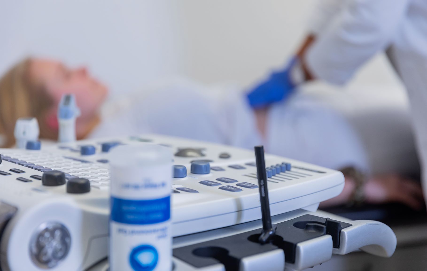
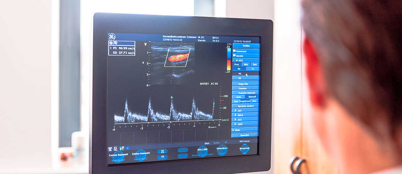
ABI measurement
The measurement of the ankle-brachial index (ABI) serves for early detection of peripheral arterial disease, which affects the arteries of the arms and legs. With the help of a simple and quickly feasible Doppler ultrasound examination, we measure the blood flow in the extremity arteries and calculate the ABI ratio. If the calculated ABI ratio is significantly reduced, the vessels in the legs are likely to be damaged, indicating arterial occlusive disease. Early detection is important because this disease is often well advanced before it causes symptoms.
The measurement of the ankle-brachial index (ABI) serves for early detection of peripheral arterial disease, which affects the arteries of the arms and legs. With the help of a simple and quickly feasible Doppler ultrasound examination, we measure the blood flow in the extremity arteries and calculate the ABI ratio. If the calculated ABI ratio is significantly reduced, the vessels in the legs are likely to be damaged, indicating arterial occlusive disease. Early detection is important because this disease is often well advanced before it causes symptoms.
We recommend an ABI measurement for patients with:
- high blood pressure
- elevated blood lipid levels
- family history of peripheral arterial disease
- older patients as a regular screening examination
Measurement of the pulse wave velocity (PWV)
Pulse wave analysis (PWA) is used to measure vascular stiffness. Changes in the arterial wall can be detected through PWA, which occur in the early stages of atherosclerosis. A decrease in vascular elasticity, as a precursor to atherosclerosis, can be detected even before a manifest underlying disease is present, and is therefore suitable for estimating the future risk of atherosclerosis. In case of elevated values, preventive measures would include avoiding high blood pressure.
The measurement takes about 5 minutes and is painless.
Pulse wave analysis is recommended for diseases that favor atherosclerosis or in which a change in pulse wave velocity may be present, including:
- Obesity
- Arterial hypertension (high blood pressure)
- Chronic kidney insufficiency (chronic renal failure)
- Diabetes mellitus (sugar disease)
- Hypercholesterolemia (disorder of fat metabolism, which leads to elevated cholesterol levels and increases the risk of cardiovascular disease)
- Hyperuricemia (gout)
- Coronary heart disease (CHD; coronary artery disease)
- Heavy smoking
Measurement of the heart rate variability (HRV)
The frequency of heartbeats during a certain measurement period is referred to as heart rate. While heart rate describes how much the circulatory system is stressed, heart rate variability indicates how well the cardiovascular system copes with stress. The heart rate is higher during exertion than in a resting state. In addition, the heart needs time to adapt to rest or stress. Even at rest, the heart never beats completely evenly. The variability of the heart rate indicates how the heartbeat responds to various internal or external factors.
To measure heart rate variability, the heartbeat is recorded as part of an electrocardiography (ECG). This is done over a short period of 5 minutes. Fitness trackers, smartwatches, and sports watches are generally not suitable for measuring heart rate variability because they measure only the pulse, not the heartbeat.
Color Doppler ultrasonography ➤

More services
Emergency diagnostics
Emergency diagnostics
Metabolic Balance

Metabolic Balance
Pulmonary function tests

Pulmonary function tests
Check-Up

Check-Up
Vaccination and travel vaccination consultation

Vaccination and travel vaccination consultation
Ultrasound examinations
Ultrasound examinations
Skin cancer screening

Skin cancer screening
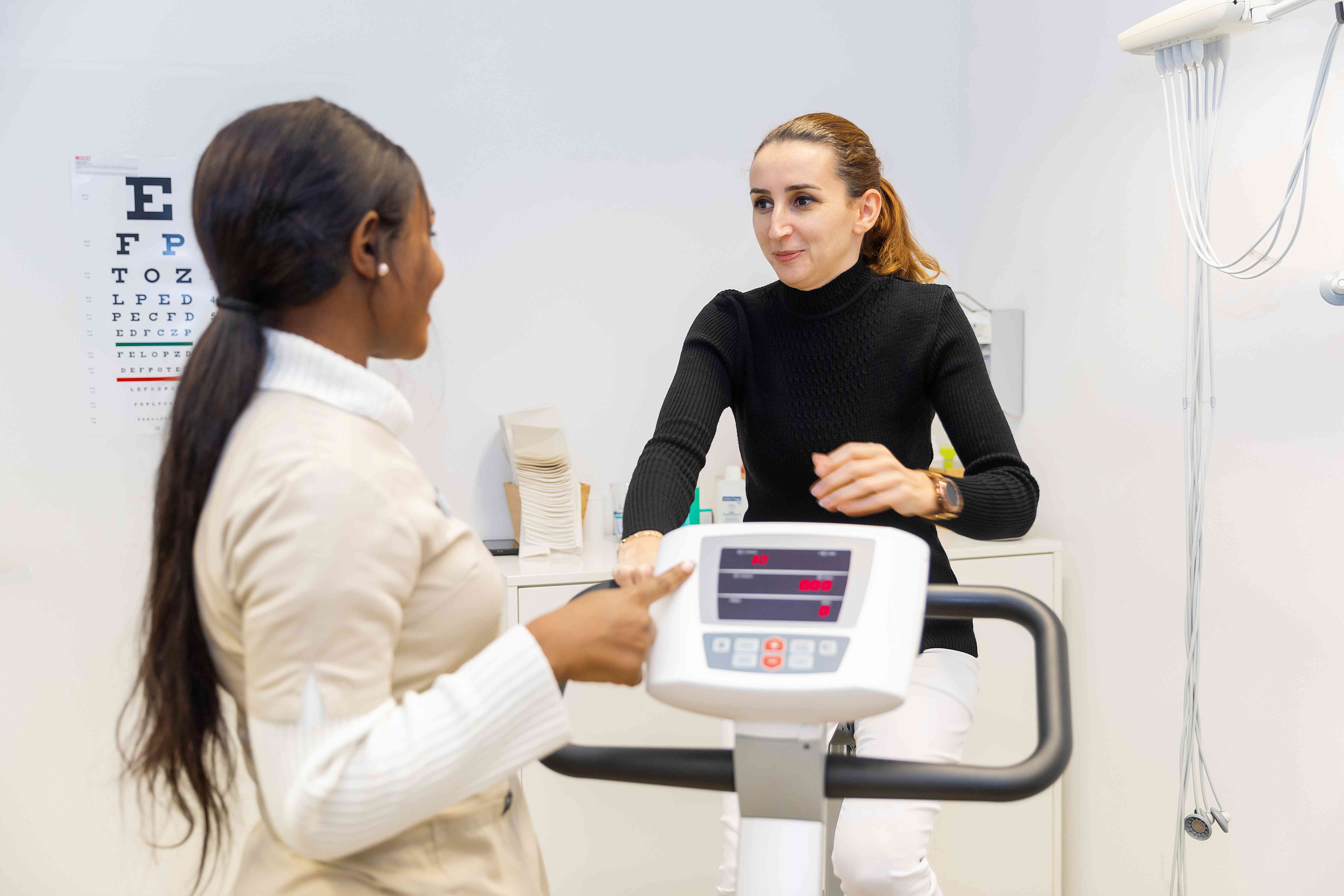
Body Fat Measurement

Body Fat Measurement
Color Doppler ultrasonography
Color Doppler ultrasonography
Stress-ECG
Stress-ECG
Laboratory tests
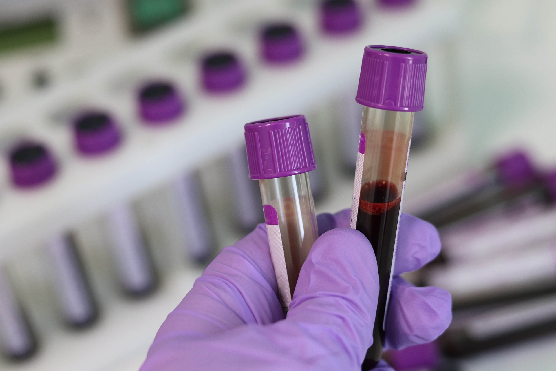
Laboratory tests
Cardisiography

Cardisiography
DMP (Disease management program)

DMP (Disease management program)
Cancer screening

Cancer screening
Individual Health Services (IGeL)

Individual Health Services (IGeL)
Cardiological examinations
Cardiological examinations