


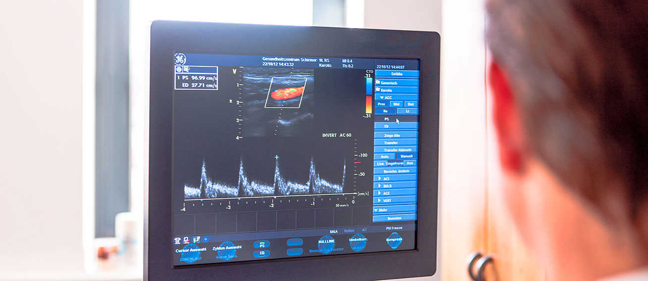
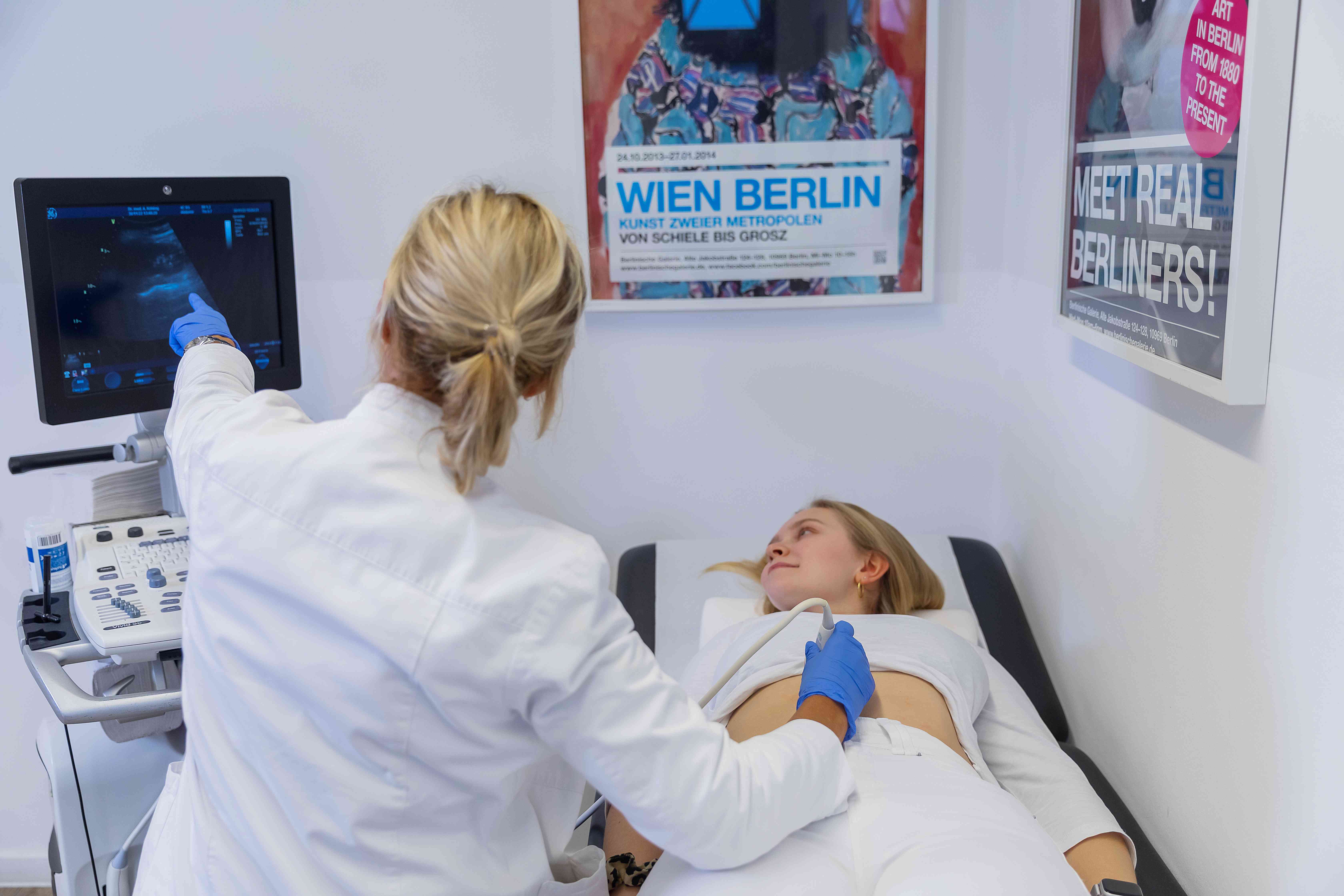
Color Doppler ultrasonography
Duplex ultrasonography is often the first choice for examining blood vessels. This is because the effects of narrowing on blood flow can be assessed in "real-time."
In the diagnosis of vascular diseases, duplex ultrasonography is a simple, painless, and risk-free examination. Using ultrasound, constrictions caused by arterial calcification, arterial expansions (so-called aneurysms), vascular injuries, and congenital vascular malformations (e.g. AV fistulas) can be visualized. More complex examination methods, such as X-rays, CT, or MRI, often become unnecessary.
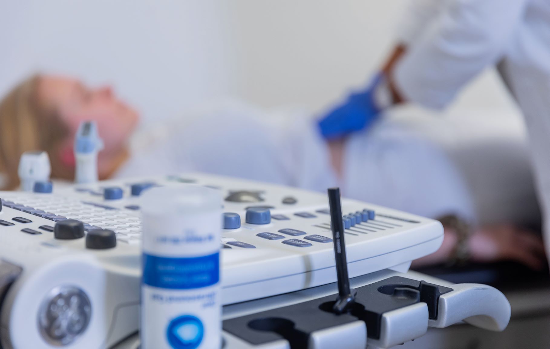
How does duplex ultrasonography work?
Like any other ultrasound examination, duplex ultrasonography uses ultrasound waves that are reflected to varying degrees depending on the structure and density differences of the targeted organs. The device recaptures the reflected sound waves through the transducer. Based on the data, an image of the internal organs and other body structures is created on a monitor. In addition, duplex ultrasonography also makes the blood flow in the vessels and organs visible and audible and determines its speed.
Like any other ultrasound examination, duplex ultrasonography uses ultrasound waves that are reflected to varying degrees depending on the structure and density differences of the targeted organs. The device recaptures the reflected sound waves through the transducer. Based on the data, an image of the internal organs and other body structures is created on a monitor. In addition, duplex ultrasonography also makes the blood flow in the vessels and organs visible and audible and determines its speed.
Applications of duplex ultrasonography:
- Vascular narrowing
- Early detection of arterial calcification
- Thrombosis diagnostics
- Varicose vein disorders
- Postoperative monitoring or stent implantation checks
- Pathological vascular expansions
- Vascular malformations
- Inflammatory vascular diseases (e.g., rheumatic diseases)
- Heart diseases

More services
Emergency diagnostics
Emergency diagnostics
Pulmonary function tests

Pulmonary function tests
Cardisiography

Cardisiography
Vaccination and travel vaccination consultation

Vaccination and travel vaccination consultation
Laboratory tests
Laboratory tests
Individual Health Services (IGeL)

Individual Health Services (IGeL)
Cardiological examinations
Cardiological examinations
Check-Up
Check-Up
Color Doppler ultrasonography
Color Doppler ultrasonography
Skin cancer screening

Skin cancer screening
DMP (Disease management program)

DMP (Disease management program)
Ultrasound examinations
Ultrasound examinations
Metabolic Balance

Metabolic Balance
Body Fat Measurement

Body Fat Measurement
Cancer screening

Cancer screening
Stress-ECG
Stress-ECG